X-Ray, CT Scan & MRI: Peek Inside Your Body Like a Detective
Ever wondered how your bones, muscles and organs appeared before they cut you open? That is precisely what X-rays, CT scans and MRIs do: they enable medical professionals to take a peek inside your body and act like detective agents in the future.
However, here is the twist: every scan narrates an entirely different story. One gives you shadows, another cuts you into pieces and the last gives you a masterpiece on soft tissue. By the time you get to the bottom of this blog post, you will get to know how each scan functions, what it displays, and also how you can be able to see just the fundamentals on an X-Ray like a mini detective.
The Magic of X-Rays: Shadows in your body.
We will begin with the easiest but most common scan: the X-ray. Think of it as a black and white picture of your inner world. X-rays are based on electromagnetic radiation, which is a form of energy that may penetrate your body. The trick to it is that X-rays are absorbed differently in various tissues.
Most of the radiations are attached to dense tissue, such as bones, which would be seen to be white in the image. Those that are less dense such as muscles and organs will be seen as grey colors and the spaces filled by air such as your lungs are black.
The reason why doctors prefer X-rays is that they are fast, cheap, and readily available, and thus most cases, they are used. You must have seen an X-ray, either when you got your bone broken, had a chest infection, or even when you went to see the dentist.
They are ideal to identify fractures, dislocations of the joints or items that have been swallowed. You can find out some rudimentary ways of reading an X-ray even when you are not a radiologist.
E.g., consider the bones: they must be smooth and continuous. A fracture could be witnessed in any cracks or abnormal lines.
Examine joint space- irregular spacing may also indicate cartilage injury or swelling.
The appearance of your lungs should be dark and clear; the appearance of white areas could be fluid or infection. The heart and other organs must be proportionate in their shapes and metallic objects such as coins or implants should be seen prominently.
Imagine that X-rays are shadow maps – patterns and shapes that do not give the entire story. The limitation? X-rays are insensitive to soft tissues. In those places, ligaments, tendons, and nerves hardly appear, and in that case, a doctor will require something more intense in case of injury.
CT Scans: Cutting You into Slices.
Input the CT scan – short computer tomography. In case X-rays are snapshots, CT scans are 3D maps constructed out of hundreds of slices. In the technology, rotating beams of X-rays are used to obtain images from various angles, which are then restructured by a computer into cross-sections of your body in great detail.
Picture a loaf of bread. When it is cut into thin layers, you can observe the interior structure and texture. That is what a CT scan does on your body. It can depict bones, organs and soft tissue in great details, hence doctors tend to use CT scans in case of an emergency or when X-ray does not give enough information.
CT scans are good in the detection of internal bleeding, organ trauma, kidney stones, brain haemorrhages, tumors, and complications of the chest. They are the referral of the trauma patients or in any case of unplanned, inexplicable pain. CT scans prove to be a savior in hospitals due to their speed and precision.
The CT scans however, consume more radiation compared to conventional X-Ray, hence the physicians balance the information requirement with exposure. They work faster than MRI, yet are not as soft as radiation is concerned.
MRI: The Masterpiece in Soft Tissues.
However now, when X-rays and CT are photos and slices, MRI (Magnetic Resonance Imaging) is a high-definition portrait of your soft tissues. It doesn’t use radiation. MRI on the other hand, involves powerful magnetic fields and radio waves to orient the hydrogen atoms in your body.
The release of this energy by these atoms leads to the creation of very detailed images by the computer.
MRI is superior in imaging of soft tissues such as the brain, spinal cord, muscles, tendons and ligaments. It is also applicable in the identification of tumors or internal inflammation that may not be identified in other scans.
Compared to X-Ray or CT, MRI is slow and it makes you lie in a noisy machine but what comes there is an amazingly detailed picture.
Physicians prefer MRI in cases when they have to examine chronic pain, neurological disorders, ligament or tendon trauma, or tumours of soft tissues. It does not harm most of the people but persons having some metal implants, pacemakers and shrapnel in their body must either avoid MRI or take special precautions.
Why do Physicians prefer one Scan over the other?
One can be tempted to believe that the strongest scan is the best one, but that is not always the case. Physicians make decisions depending on what they have to see. The initial X-rays are used for the bones and general injury.
The quick emergency method is CT scans which reveal the organs and bones simultaneously. MRI is the comprehensive expert of soft tissues and reveals those that are not discovered by X-Ray and CT.
This logic enables the patients to feel more educated and less intimidated. Every scan is a kind of detective: one sees the shadows, another cuts the body into layers, the last one takes the soft tissues with such lovely clarity with its camera.
Common Myths About Scans
There are several myths about these scans:
- CT and MRI are identical: No. CT involves the use of X-ray radiation and MRI involves the use of magnets.
- MRI is harmful: MRI is harmless to the majority of people; some metal implants should be taken care of.
- X-Ray knows it all: Bones, yes. Soft tissues, no.
- CT will be superior to X-Ray: CT is more elaborate and you are exposed to more radiation, hence it is only used in selective cases.
Appreciation of such myths can help to avoid the unwarranted fear and confusion.
Peeping inside the body like a detective.
Being familiar with what scans are, the ability to read and interpret an X-Ray brings an extra dimension of curiosity. Although you are not to diagnose yourself, it is rather entertaining to identify easy patterns. Find smooth bones, equal joint spaces, black lungs, and equal organs. Identify metallic objects or strange shadows. It is a kind of a black and white journey to another world where a visit to the hospital becomes a learning process.
CT scans and MRI may be harder to interpret for a layperson, but appreciating their purpose is enough. Imagine a CT as slicing your body into ultra-thin layers and MRI as painting a detailed portrait of your muscles, nerves, and organs — you can almost see what the doctor sees.
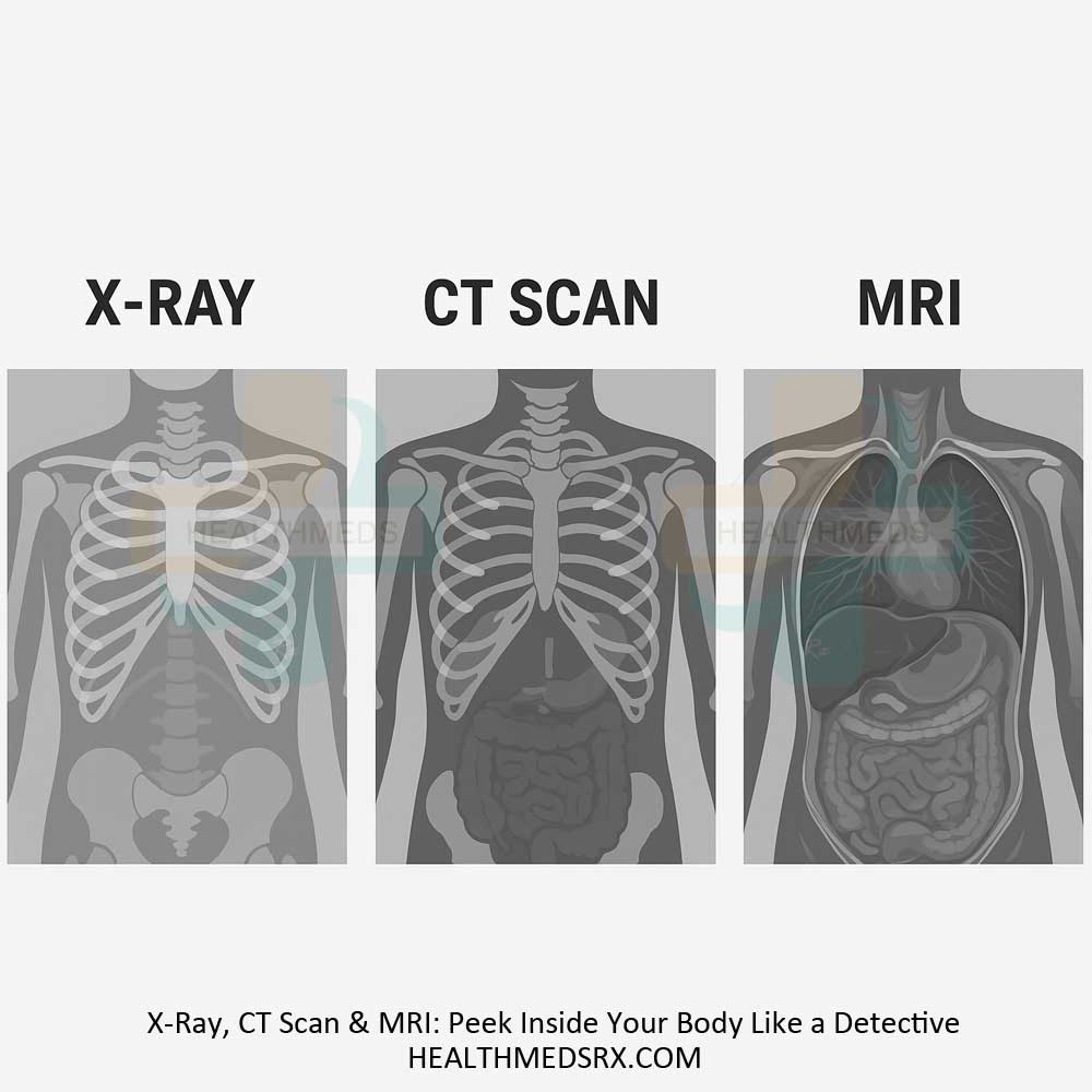
The difference: X-Rays, CT scans, and MRIs
X-Rays, CT scans, and MRIs are not competitors; they’re different detectives with unique superpowers. Each one tells a story about your body that the others can’t:
- X-Ray: Quick shadows and basic structure
- CT Scan: Layers revealing organs and bones
- MRI: Soft tissue masterpieces with no radiation